Announcing: 2026 Signals that Move Us Image Competition Winners
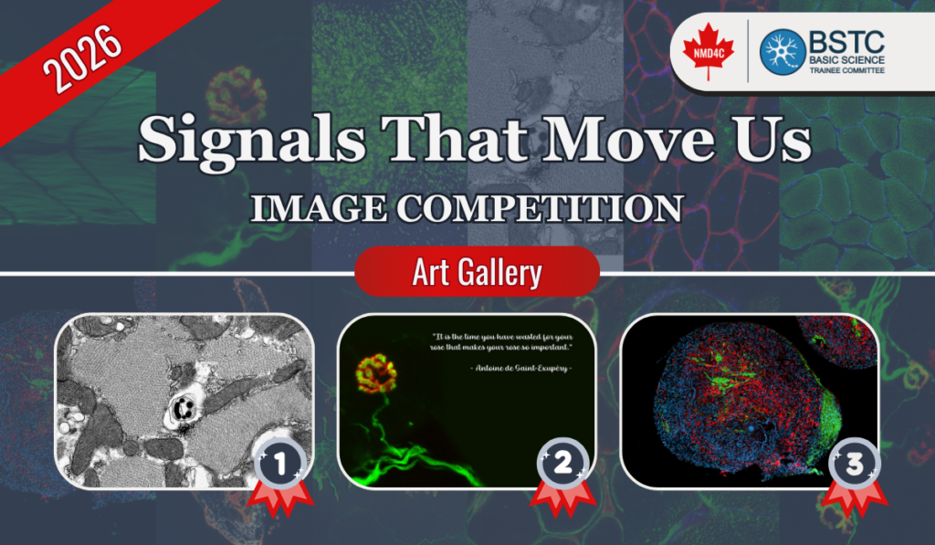
Research often uncovers incredible visual beauty alongside data and discovery.
This year, our community highlighted that intersection through the Signals That Move Us Image Competition. We received 12 entries from our trainees, each offering a unique look at the signals, structures, and cellular dynamics driving our research. With so many strong entries, selecting the top spots was a difficult task for the committee. We want to extend a sincere thank you to everyone who took the time to submit their work and share their microscopy and scientific
imaging with the community.
Meet the Winners

1st Place: Madison Garibotti
PhD Candidate, York University
Supervised by Dr. Christopher Perry
Image Title: You Can Find Happiness in the Oddest Places!
2nd Place: Dr. Tatiana Koike
Postdoctoral Research Fellow, CHU Sainte Justine and Université de Montréal
Supervised by Dr. Nicolas Dumont
Image Title: The Neuromuscular Junction as a Living Rose

3rd Place: Alexa Derksen
PhD Candidate, University of Ottawa and CHEO Research Institute
Supervised by Dr. Hanns Lochmuller
Image Title: 3D iPSC derived Neuromuscular Organoid

Image Competition Art Gallery now live!
In addition to the top three winners, we received many outstanding submissions from neuromuscular trainees across Canada. The full exhibition is now live. Our online art gallery showcases all 12 submitted images, including the top three, featuring work that ranges from cellular staining to complex structural patterns.
Explore the Art Gallery
Planning for Next Year competition
To all of our trainees: as you collect your data over the coming months, keep an eye out for
striking visuals. Whether you are working with confocal microscopy, western blots, or
computational models, we encourage you to save your best shots. We look forward to seeing your submissions for next year’s competition. Congratulations again to our winners, and thank you to everyone who participated.
Read next...
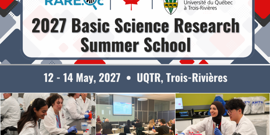
2027 NMD4C–RARE.Qc Basic Science Research Summer School Applications Are Now Open!
Deadline: December 1, 2026
2027 NMD4C–RARE.Qc Basic Science Research Summer School Applications Are Now Open!
The summer school offers neuromuscular and rare disease research trainees the opportunity to participate in an intensive three-day training program focused on cutting-edge experimental and translational research techniques.
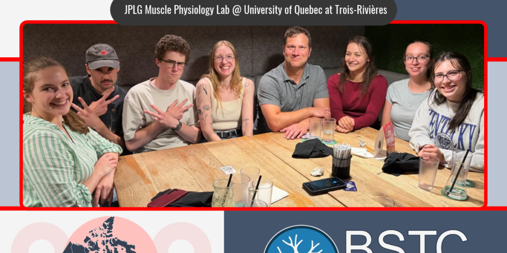
BSTC: Lab of the Month – Dr. Jean-Philippe Leduc-Gaudet’s Muscle Physiology Lab at UQTR
Our new Lab of the Month series highlights members of the NMD4C community and the diverse research, expertise and collaborations taking place across our network. Each month, we’ll introduce a different lab, meet the people behind the research and learn more about the work they are advancing. Kicking off the series, we’re featuring the lab…